From Cloudy to Clear: What Actually Happens During Cataract Surgery?
Vision is often taken for granted—until it begins to disappear.
For people with cataracts, sight doesn’t vanish suddenly.
Instead, the world gradually dims, colors lose their vibrancy, and sharp edges soften into a permanent blur.

This slow decline is caused by a deceptively small structure deep inside the eye: the crystalline lens.
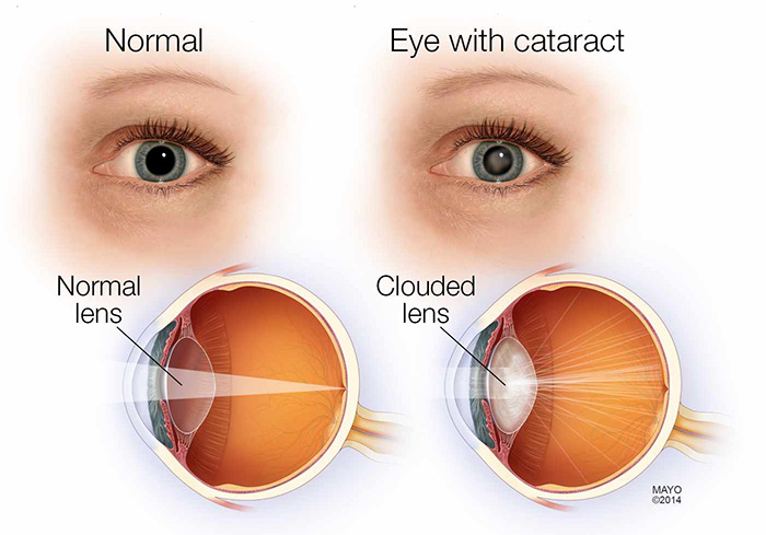
In a healthy eye, the lens is perfectly transparent, bending incoming light so it focuses sharply on the retina.
Over time, however, the proteins that make up this lens begin to break down.
Aging, injury, diabetes, medications, or prolonged UV exposure can cause these proteins to clump together, turning the lens yellow and opaque.
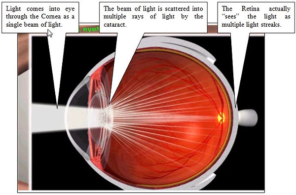
This is a cataract—essentially a biological fog that scatters light instead of focusing it.

As the cataract thickens, light struggles to pᴀss through.
Vision becomes dull, night driving grows difficult, and glare from lights becomes overwhelming.
Eventually, glᴀsses can no longer compensate.
At this point, surgery becomes the only solution—and remarkably, one of the safest and most successful surgeries in modern medicine.
Before surgery, ophthalmologists ᴀssess the cataract using a slit-lamp microscope.

By shining a narrow beam of light through the eye, they can visualize exactly how dense the cloudy lens has become.
This detailed mapping allows surgeons to plan the procedure with microscopic precision.
On surgery day, the process is swift and surprisingly gentle.
The patient remains awake but comfortable.
Anesthetic eye drops numb the surface, eliminating pain while allowing awareness.
A small device called a speculum gently holds the eyelids open, preventing blinking during the operation.
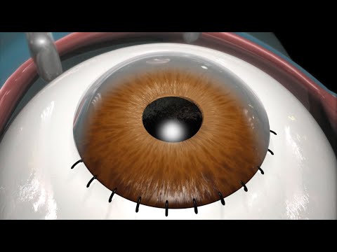
The surgeon begins by creating a tiny incision—about 2 millimeters wide—at the edge of the cornea using a precision blade.
This opening is so small that it is self-sealing and rarely requires sтιтches.
Through this microscopic gateway, all surgical instruments will pᴀss.
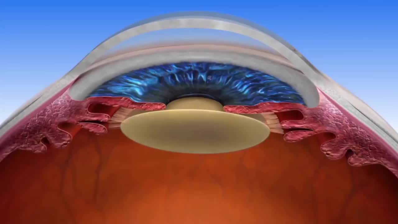
Next comes one of the most critical steps: accessing the lens while preserving its natural housing.
The lens sits inside a thin, transparent capsule.
Using delicate instruments, the surgeon carefully removes a circular opening from the front of this capsule in a step known as capsulorhexis.
This must be smooth and precise, as the remaining capsule will later support the new artificial lens.
To loosen the cataract, a gentle wave of sterile fluid is injected beneath the capsule.
This process, called hydrodissection, separates the hardened lens from its surrounding structure, allowing it to move freely.

This makes removal safer and more controlled.
The heart of the surgery is a technique called phacoemulsification.
A tiny ultrasonic probe enters the eye and vibrates at extremely high frequencies.
These vibrations shatter the hardened lens into microscopic fragments.
Simultaneously, a built-in vacuum system suctions the fragments out, clearing the eye of the cloudy material that once blocked vision.
Once the dense cataract is gone, softer residual fibers are carefully removed using irrigation and aspiration tools.
The surgeon meticulously cleans the inside of the capsule, polishing it until it becomes clear and pristine—ready for its replacement.
That replacement is an intraocular lens, or IOL.
Custom-selected for the patient’s eye, this artificial lens is made from advanced biocompatible materials.
It is folded тιԍнтly and inserted through the same tiny incision.

Once inside the eye, it gently unfolds, its flexible arms—called haptics—anchoring it perfectly within the capsule behind the pupil.
With the new lens in position, the eye is re-pressurized with sterile fluid.
The small corneal incision seals naturally, held closed by the eye’s internal pressure.
No sтιтches.
No dramatic recovery.

Instantly, the eye’s optical system is restored.
Light now travels freely through the clear cornea and artificial lens, focusing sharply onto the retina.
The brain receives crisp, bright images once again.
Recovery happens quickly.
On a cellular level, corneal surface cells slide over the incision within hours, sealing it completely.
Mild inflammation subsides within days.
For many patients, vision improves within 24 hours, with colors appearing noticeably brighter—often described as seeing the world in high definition after years of fog.
Cataract surgery is more than a medical procedure.
It is a triumph of precision engineering, biology, and technology working together.
In less than 30 minutes, a lifetime of fading vision can be reversed—restoring independence, confidence, and clarity to millions of people every year.


